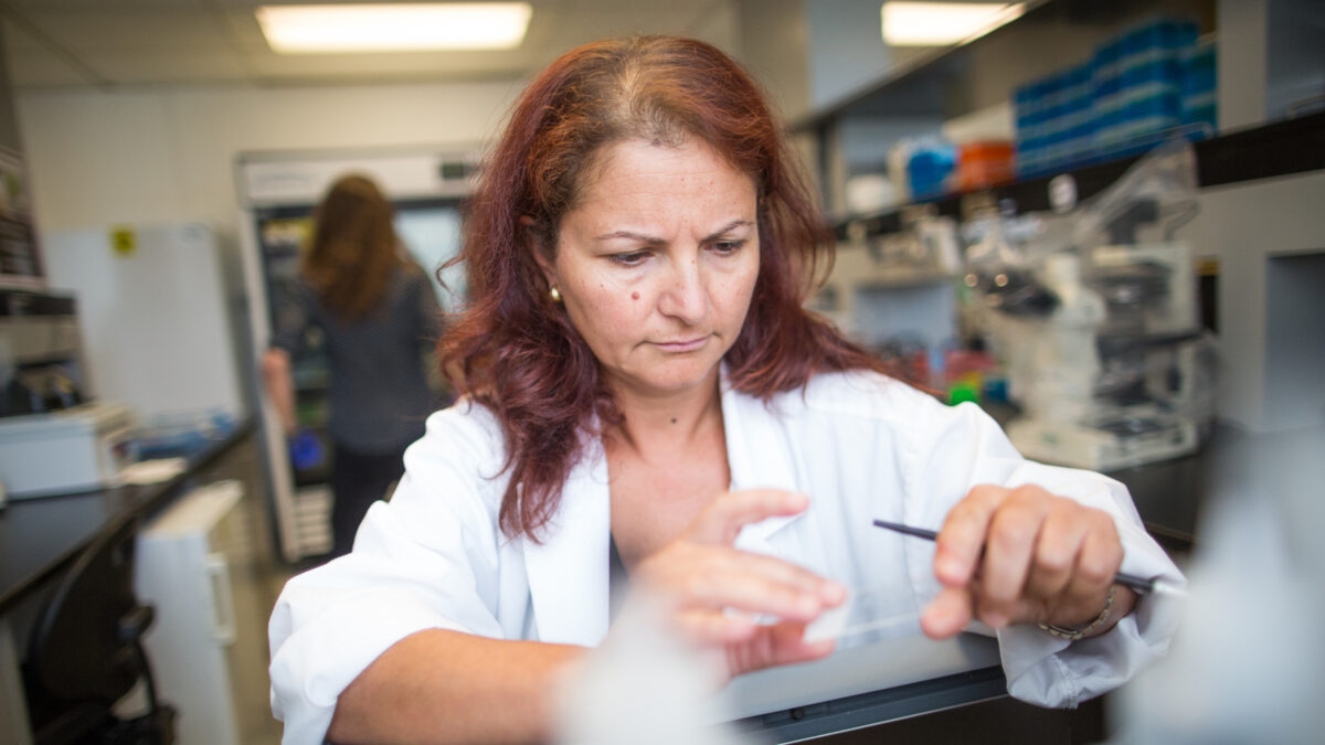
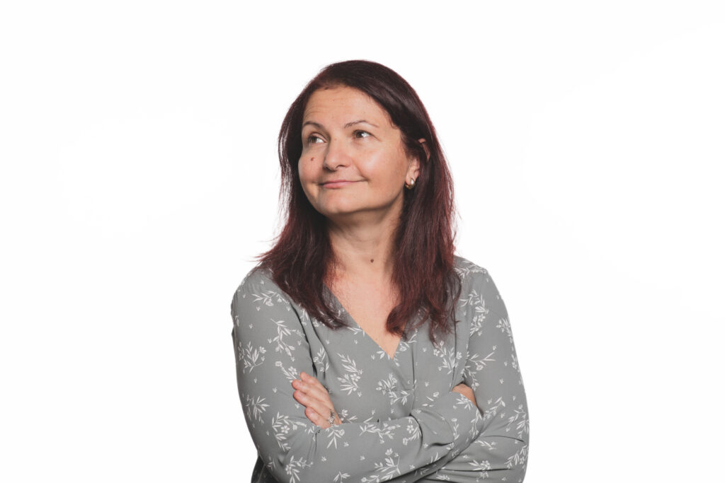
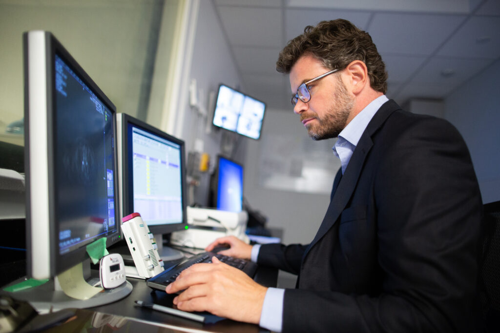
Cardiologist Dr. Phyllis Billia is investigating whether the heart can be altered so it’s able to regenerate.
Tim Fraser
Pushing the boundaries of scientific discovery
Philosophers and poets will muse about the human heart; at the Peter Munk Cardiac Centre, world-class researchers are coming closer to discovering what makes it tick.
Can a damaged heart learn to fix itself? There’s the question Dr. Phyllis Billia is looking to answer through her pioneering research at University Health Network’s Peter Munk Cardiac Centre. Dr. Billia, cardiologist, Peter Munk Cardiac Centre Research Lead and Co-Director of the Peter Munk Cardiac Centre Cardiovascular Biobank, says part of the answer may rest in how hearts develop in the first place.
When a baby is born, the heart “proliferates,” or grows, for a period of time. Then it stabilizes and stops growing, she explains.
But what if a damaged heart could regrow?
“How can we coax it back?” Dr. Billia asks.
The problem is that the heart is “not really a great regeneration organ,” she notes. When a surgeon opens up an artery that caused a heart attack, there is usually irreparable damage. But Dr. Billia is investigating whether the heart could be altered so that it’s able to regenerate.
“If we could get viable heart tissue to repair the damage, then we could prevent heart failure in the kinds of patients I see all the time,” she says.
The key to a self-repairing heart may be delivering a drug that could induce neighbouring cells to reduce the damage, Dr. Billia explains. A heart cell’s ability to regenerate is suppressed by natural “roadblocks,” so they are seeking a drug that can remove those roadblocks. Dr. Billia’s team has figured out the molecular “signatures” in the early stages of what could drive this potential repair. They’ve also done modelling where they have removed certain genes from the mix, to see what changes.
“If we could get viable heart tissue to repair the damage, then we could prevent heart failure.”
Dr. Phyllis Billia
163,000
The number of patient visits at the Peter Munk Cardiac Centre per year, performing everything from routine examinations to the most complex surgeries and transplants.
The team is using cancer research as a model, says Dr. Billia, citing how some stem cell research investigates how to encourage healthy cells to replicate instead of cancerous ones.
“We’re looking at how we can push the heart into ‘cell-cycle’ like that. The next stage is to use drugs to target whether we can induce repair after we open an artery,” she says.
“Really, no one else is doing what we’re doing in the way that we’re looking at regeneration [in cardiovascular research].”
Dr. Billia’s work is very much in keeping with the spirit of the centre’s founder and benefactor, the late Peter Munk.
“He was incredible – thoughtful and humble, yet quite visionary in terms of knowing what to focus on in offering his support. I consider him a man with a lot of foresight,” she says.
An ‘incredible, exciting’ time in research
The Peter Munk Cardiac Centre treats some 163,000 patients a year, performing everything from routine examinations to the most complex surgeries and transplants. That’s only part of the job though. While healing present-day patients, the centre is also keenly focused on looking ahead to the future of cardiovascular care.
The physicians and researchers at this world-class institution want to know why: Why are some people born with heart disease? Why do people develop aneurysms? Why do some patients recover from heart attacks while others do not? These researchers are seeking – and finding –new ways to prevent, treat and understand heart disease.
“We have multiple initiatives on the go,” says Dr. Barry Rubin, vascular surgeon and the Peter Munk Cardiac Centre’s Medical Director and Chair. “It’s an incredible, exciting time.”
The Peter Munk Cardiac Centre is well into what Dr. Rubin calls its Discovery Program – ongoing research and breakthroughs that go back more than 80 years at Toronto General Hospital and University Health Network, before the Peter Munk Cardiac Centre was established in 1993 through donations led by the late Peter Munk and his wife, Melanie.
“We have multiple initiatives on the go. It’s an incredible, exciting time.”
Dr. Barry Rubin
284,000+
The number of samples collected and stored in the Peter Munk Cardiac Centre Cardiovascular Biobank.
For example, researchers are mapping single cells in unprecedented detail and exploring the potential of stem cells to support heart repair. They are using ever-more detailed imaging to treat patients with less intervention and more precision. And they’re making strides in areas ranging from immunology to genetics.
This is the future of discovery at the Peter Munk Cardiac Centre and the world-class researchers who are driving it.
Probing the mysteries of genetics
Could doctors predict who will develop heart disease and who will not?
While environment and lifestyle are always factors, some heart disease is genetic and inherited, says Dr. Raymond Kim, Scientific Lead of the Cardiac Genome Project at the Ted Rogers Centre for Heart Research, which spans the Peter Munk Cardiac Centre, The Hospital for Sick Children and the University of Toronto.
“Naturally, those families with a history of heart disease would want genetic testing for the one particular gene that may be the cause,” Dr. Kim says.
But up until about five years ago, that kind of genetic testing was not easily available because of a hefty cost. It would cost about $1,000 per gene and take about three months for the results to come back. An unrealistic proposition, since there are upwards of 25,000 genes in the human genome.
“The breakthrough is that now, testing all 20,000–25,000 genes – not just one – costs just $2,000 and takes about three months for everything,” Dr. Kim says. “It took 20 years and $3-billion to map the human genome. Now we have terabytes of information.”
20,000+
The number of genes the Cardiac Genome Project at the Ted Rogers Centre for Heart Research is able to test for just $2,000, when previously it would have cost $1,000 per gene.
Dr. Kim says he and his team are now using that information, with the goal of finding out more about the relationship between genetic disorders and heart disease. Ultimately, those relationships could lead to doctors being able to gain more insight into who will develop heart disease in future.
“Heart disease has one of the highest yields for new discover, so it’s a good place to be.”
Dr. Raymond Kim
The biggest obstacle to the next phase of this discovery breakthrough is not technology, Dr. Kim adds. “It’s computational power, and our understanding of the actual biology of the genes.”
Despite the challenges ahead, Dr. Kim has high hopes for his field. “Heart disease has one of the highest yields for new discovery, so it’s a good place to be,” he says.
Understanding aneurysms
“If you have an aneurysm, there’s a 50 per cent chance you’ll be dead before you even reach the hospital,” says Dr. Clinton Robbins, scientist and the Peter Munk Chair in Complex Aortic Therapy.
An aneurysm – which occurs when an artery wall is weakened and balloons out – can lead to a blood vessel or heart rupture and sudden death.
Dr. Robbins is on the hunt for what causes these frequently fatal occurrences and how to prevent them, including how they might be triggered by cigarette smoke. He says they know that the majority of aneurysms can’t be explained by genetic predisposition, so they’re looking at other risk factors, such as the development of plaque buildup in the arteries (atherosclerosis).
“There’s a lot of anecdotal information, but we’re trying to do better than that,” says Dr. Robbins.
Making these kinds of connections could help scientists determine who is at risk for aneurysms and even help prevent them from happening.
“We’re the largest centre for repair [of aneurysms] in Canada, but treatment can be complicated.”
Dr. Clinton Robbins
500
Out of 500 aortic aneurysm repairs performed annually at the Peter Munk Cardiac Centre, 190 are complex and 150 have open chest surgical repair and 40 of the complex cases are minimally invasive, the highest volume in Canada.
Dr. Kong Teng Tan, interventional radiologist and division head of interventional radiology at the Peter Munk Cardiac Centre, says they are also looking at how to minimize invasive treatment for aneurysms.
“We’re the largest centre for repair [of aneurysms] in Canada, but treatment can be complicated,” he says.
Right now, aneurysms are most commonly treated with stents – tubes reinforced with wire mesh that are inserted into weakened blood vessels. That’s current technology. In the future, Dr. Tan expects that his research will lead to smaller and smaller stents – and perhaps no stents at all.
“We’ve been using stents for years, but there are limits to what they can do. In five or six years, there will be better designs for stents, [but] we’re looking at even finer technology. For example, we could use stem cells to rebuild blood vessels, instead of using a stent.”
The idea is that blood would be taken from the patient and filtered. Once the specific “signalling factors” that stimulate circulation in the blood vessels were identified, they could be injected back into the patient to help rebuild the artery.
“There are already clinical trials underway,” says Dr. Tan of this novel approach.
The centre also has new imaging equipment that will enable them to monitor how blood vessels are behaving, a great benefit to this sort of investigation, he adds.
“PET [positron emission tomography] scanners can let us see whether the stem cells that are injected are stimulating the blood vessels.”
Predicting patients’ cardiovascular health
For Dr. Patrick Veit-Haibach, the future of discovery is in imaging biomarkers.
“In the coming years, I would hope that the projects we’re working on will help us and patients make decisions on transplants and treatments,” says Dr. Veit-Haibach, a radiologist and nuclear medicine physician who came to the Peter Munk Cardiac Centre from the University of Zurich in 2017. “For example, we could use imaging biomarkers to predict the success of a procedure, as well as when it is important to intervene earlier.”
A biomarker is any measurable biological characteristic that can measure a disease state or bodily process. Researchers can use sophisticated imaging tools, such as the centre’s cutting-edge PET-MRI (positron emission tomography-magnetic resonance imaging) scanner to identify biomarkers and measure disease activity.
Dr. Veit-Halbach is working on a number of complex projects that involve mapping the heart and how it functions. Ultimately, they all come down to answering simple questions: Why do certain things happen in the heart, and what can be done?
“We’ll look at patients with treated cancer, for example, who also have shortness of breath,” he says. “We want to try to figure out through imaging what the underlying reasons are. Perhaps it’s cardiac dysfunction, perhaps lung hypertension.”
“In the coming years, I would hope that the projects we’re working on will help us and patients make decisions on transplants and treatments.”
Dr. Patrick Veit-Haibach
Dr. Veit-Haibach and his co-researchers are also looking at the metabolism of heart attacks, using imaging to figure out why some patients who have heart attacks might recover, while others don’t survive or have limited recovery.
“Another project is with patients [who have received] radiation therapy to the neck,” Dr. Veit-Haibach adds. “Those patients are known to have higher risks for cardiovascular events; however, nobody knows the exact reason.”
He and other experts suspect that the arteries supplying the brain are altered after the radiation therapy (also called radiotherapy).
PET-MRI scans can look at those vessels in astonishing detail, pinpointing areas of very subtle inflammation that were never detectable before.
“We can compare what we see to ‘normal’ vessels, because not all vessels [would have been] in the radiotherapy field. We hope to see biomarkers that will determine the difference,” Dr. Veit-Haibach says.
The centre’s infrastructure and funding enable Dr. Veit-Haibach to work toward the future of discovery with the confidence that there will be more breakthroughs. The PET-MRI equipment is a boon, and so is the positive atmosphere at the Peter Munk Cardiac Centre, he says.
“It’s very collaborative,” he says. “I have been in places where it’s much more complicated.”
Related Stories